Female Reproductive Organs Diagram : Female Reproductive System With Labelled Diagram Presentation Graphics Presentation Powerpoint Example Slide Templates - The female reproductive system has two functions:
byAdmin-
0
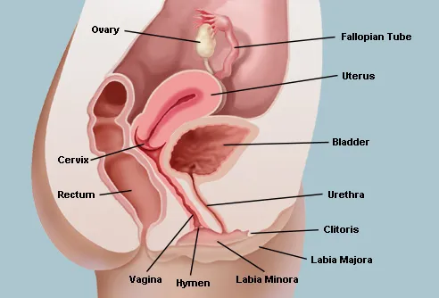
Female Reproductive Organs Diagram : Female Reproductive System With Labelled Diagram Presentation Graphics Presentation Powerpoint Example Slide Templates - The female reproductive system has two functions:. They produce gametes and help in the secretion of hormones, which regulates the gamete formation and secondary sexual characters in females. The broad ligament is a reflection from the peritoneum. The major organs of the female reproductive system include: A female reproductive organ, often paired, that produces ova and in mammals secretes the hormones estrogen and progesterone. This diagram depicts picture of female reproductive system diagram 1024×1204 with parts and labels.
Label the diagram and write the function of each part below. It includes the mons, clitoris, labia, hymen and the opening of the urethra (picture 3). Undifferentiated male female gonad testis ovary mullerian duct appendix testis fallopian tubes mullerian duct prostatic utricle uterus, proximal wolffian duct rete testis rete ovarii mesonephric tubules efferent ducts epoophoron Many girls and teens wonder about their reproductive organs and whether they are normal. Start studying female reproductive organs.
Female Reproductive System Organs Function And More from img.webmd.com Labeled diagram of the external female reproductive organs the perineum is the region where the pelvic floor muscles are located. The female reproductive system is responsible for producing the female gamete, also known as the ovum or egg.in this post, we will tell you about the different parts of the female reproductive system, their functions, and some interesting facts about this organ system. Start studying female reproductive organs. Female reproductive system physiology the reproductive cycle. Females make millions of eggs every day. The information below provides the name for each body part and its specific function, along with a diagram to show where it is located on the female body. Female anatomy includes the external genitals, or the vulva, and the internal reproductive organs. The function of the external female reproductive structures (the genital) is twofold:
The broad ligament is the inclusive term for the mesovarium, mesosalpinx, and mesometrium that suspend the ovary, uterine tubes, and uterus, respectively, from the dorsolateral wall of the sublumbar region.
The major organs of the female reproductive system include: They produce gametes and help in the secretion of hormones, which regulates the gamete formation and secondary sexual characters in females. This diagram depicts picture of female reproductive system diagram 1024×1204 with parts and labels. The first is to produce egg cells, and the second is to protect and nourish the offspring until birth. Unlike the male reproductive organs, the female reproductive organs are located mostly in the pelvic cavity. Is the largest organ in the normal female pelvis when the urinary bladder is empty The female reproductive cycle is the process of producing an ovum and readying the uterus to receive a fertilized ovum to begin pregnancy. If an ovum is produced but not fertilized and implanted in the uterine wall, the reproductive cycle resets itself through menstruation. Our experts describe the functions of female reproduction, including ovulation, fertilization, and menopause. The female reproductive system has two functions: A female reproductive organ, often paired, that produces ova and in mammals secretes the hormones estrogen and progesterone. Undifferentiated male female gonad testis ovary mullerian duct appendix testis fallopian tubes mullerian duct prostatic utricle uterus, proximal wolffian duct rete testis rete ovarii mesonephric tubules efferent ducts epoophoron Females make millions of eggs every day.
Find more on the female reproductive organs, the menstrual cycle, and more. Start studying female reproductive organs. The female reproductive anatomy includes both external and internal structures. Cranial view of bovine female reproductive ortans. A duct through which an ovum passes from an ovary to the uterus or to the exterior (called fallopian tubes in humans).
9 942 Female Reproductive System Illustrations Clip Art Istock from media.istockphoto.com The information below provides the name for each body part and its specific function, along with a diagram to show where it is located on the female body. Start studying female reproductive organs. Related posts of female reproductive organ diagrams female reproductive system side view. Cranial view of bovine female reproductive ortans. The first is to produce egg cells, and the second is to protect and nourish the offspring until birth. The female reproductive cycle is the process of producing an ovum and readying the uterus to receive a fertilized ovum to begin pregnancy. Label the diagram and write the function of each part below. Many girls and teens wonder about their reproductive organs and whether they are normal.
This is also known as the pudendal cleft or the cleft of venus, after the roman goddess of love.
The function of the external female reproductive structures (the genital) is twofold: A female reproductive organ, often paired, that produces ova and in mammals secretes the hormones estrogen and progesterone. Our experts describe the functions of female reproduction, including ovulation, fertilization, and menopause. The information below provides the name for each body part and its specific function, along with a diagram to show where it is located on the female body. Learn vocabulary, terms, and more with flashcards, games, and other study tools. Related posts of female reproductive organ diagrams female reproductive system side view. The broad ligament is the inclusive term for the mesovarium, mesosalpinx, and mesometrium that suspend the ovary, uterine tubes, and uterus, respectively, from the dorsolateral wall of the sublumbar region. Undifferentiated male female gonad testis ovary mullerian duct appendix testis fallopian tubes mullerian duct prostatic utricle uterus, proximal wolffian duct rete testis rete ovarii mesonephric tubules efferent ducts epoophoron The female reproductive system has two functions: A duct through which an ovum passes from an ovary to the uterus or to the exterior (called fallopian tubes in humans). This is also known as the pudendal cleft or the cleft of venus, after the roman goddess of love. It also is known as the birth canal. The female reproductive cycle is the process of producing an ovum and readying the uterus to receive a fertilized ovum to begin pregnancy.
The female reproductive system has two functions: The major organs of the female reproductive system include: It also is known as the birth canal. Find more on the female reproductive organs, the menstrual cycle, and more. Cranial view of bovine female reproductive ortans.
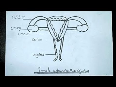
Female Reproductive System Diagram How To Draw Female Reproductive System Biology Class 10 Youtube from i.ytimg.com Learn about the female reproductive system's anatomy through diagrams and detailed facts. The first is to produce egg cells, and the second is to protect and nourish the offspring until birth. Many girls and teens wonder about their reproductive organs and whether they are normal. Ovaries are the primary reproductive organs of the female reproductive system as they directly take part in the process of reproduction. Labeled diagram of the external female reproductive organs the perineum is the region where the pelvic floor muscles are located. It includes the mons, clitoris, labia, hymen and the opening of the urethra (picture 3). This diagram depicts picture of female reproductive system diagram 1024×1204 with parts and labels. They produce gametes and help in the secretion of hormones, which regulates the gamete formation and secondary sexual characters in females.
The major organs of the female reproductive system include:
Our experts describe the functions of female reproduction, including ovulation, fertilization, and menopause. Learn vocabulary, terms, and more with flashcards, games, and other study tools. They produce gametes and help in the secretion of hormones, which regulates the gamete formation and secondary sexual characters in females. This article looks at female body parts and their functions, and it provides an interactive diagram. The female reproductive system has two functions: A female reproductive organ, often paired, that produces ova and in mammals secretes the hormones estrogen and progesterone. A duct through which an ovum passes from an ovary to the uterus or to the exterior (called fallopian tubes in humans). The major organs of the female reproductive system include: Females make millions of eggs every day. The female reproductive anatomy includes both external and internal structures. Female reproductive system side view 12 photos of the female reproductive system side view female reproductive system picture side view, female reproductive system side view labeled, female reproductive system side view parts and functions, female reproductive system side view worksheet, side view of. Female anatomy includes the external genitals, or the vulva, and the internal reproductive organs. Parts of the reproductive system.
The major parts of the reproductive system are: female organs diagram. A duct through which an ovum passes from an ovary to the uterus or to the exterior (called fallopian tubes in humans).